Correspondence between brain volumetry by magnetic resonance imaging and the Pasquier visual scale of global cortical atrophy A single-center observational study.

Main Article Content

Stalin Santiago Celi Simbaña
Andrés Javier Arroyo Salazar
Max Iván Cuesta Fernández
Paúl Javier San Martín Chung

Abstract

Introduction: The assessment of cortical atrophy is often based on subjective visual scales such as the Pasquier scale. Given the limited sensitivity of these methods, this study aims to describe the correlation between automated brain volumetry values and Pasquier scale scores in patients at Alcívar Hospital.


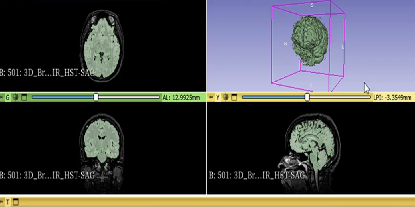
Materials and methods: This observational, retrospective study was conducted at Alcívar Hospital (Guayaquil, Ecuador) between 2023 and 2025. A total of 885 magnetic resonance imaging (MRI) scans from adults aged 65 and older were analyzed using probabilistic sampling. Variables included brain volumetry (segmented with 3D Slicer) and the Pasquier visual scale. The analysis was descriptive and employed measures of central tendency and frequencies. To mitigate bias, a structured guide of criteria and paired coding was applied to ensure data consistency and objectivity.


Results: 885 records (100% of the sample) were analyzed, with a predominance of women and of patients aged 65-75 years. According to the Pasquier scale, 66.1% presented with grade I atrophy, while grade III was observed in only 3.8%. The average global brain volume was 1,256.5 mL, with a progressive reduction as severity increased on the visual scale: 1,256.5 mL (Pasquier I), 1,228.6 mL (Pasquier II), and 1,210.5 mL (Pasquier III).


Conclusions: There is a direct correlation between the Pasquier visual assessment and quantitative volumetry. A progressive reduction of approximately 20 mL in brain volume was observed per degree of atrophy, supporting the use of volumetric measurements to objectively assess brain mass loss.

Downloads

Download data is not yet available.

Article Details

How to Cite
Correspondence between brain volumetry by magnetic resonance imaging and the Pasquier visual scale of global cortical atrophy: A single-center observational study. (2026). Actas Médicas (Ecuador), 36(1), 95-107. https://doi.org/10.61284/297
Section
Original Research
Author Biographies

Stalin Santiago Celi Simbaña, Postgrado de Imagenología, Universidad Internacional del Ecuador, Ecuador.

Médico por la Universidad Central del Ecuador (Ecuador, 2017). Médico posgradista de Imagenología por la Universidad Internacional del Ecuador, Guayaquil-Ecuador.

Andrés Javier Arroyo Salazar, Servicio de Diagnóstico por Imágenes, Hospital Alcivar, Guayaquil, Ecuador.

Médico por la Universidad de Guayaquil, (Guayaquil, 2014). Especialista en Imagenología por la Universidad de Guayaquil, (Guayaquil, 2022). Magister en Gestión Hospitalaria y Nuevas Tecnologías por la Universidad Tecnológica ECOTEC (Guayaquil, 2023). Jefe del Servicio de Radiología en el Hospital Alcívar en Guayaquil, Ecuador.

Max Iván Cuesta Fernández, Servicio de Diagnóstico por Imágenes, Hospital Alcivar, Guayaquil, Ecuador.

Doctor en Medicina y Cirugía por la Universidad de Guayaquil (Guayaquil, 2002). Especialista en Imagenología por la Universidad de Guayaquil (Guayaquil, 2010). Médico tratante del servicio de Imagenología del Hospital Alcívar.

Paúl Javier San Martín Chung, Servicio de Diagnóstico por Imágenes, Hospital Alcivar, Guayaquil, Ecuador.

Médico por la Universidad de Guayaquil (Guayaquil, 2006). Especialista en Imagenología por la Universiad de Guayaquil (Guayaquil, 2016). Magister en Gerencia y Administración de Salud por la Universidad de Guayaquil (Guayaquil, 2017).


How to Cite

Correspondence between brain volumetry by magnetic resonance imaging and the Pasquier visual scale of global cortical atrophy: A single-center observational study. (2026). Actas Médicas (Ecuador), 36(1), 95-107. https://doi.org/10.61284/297

Most read articles by the same author(s)